F1 pitstop techniques to help in resuscitation of newborn babies

On your marks...

On your marks…


Now, here’s an interesting one…

Williams has been assisting the neonatal unit at the University Hospital of Wales (UHW) in Cardiff by bringing Formula One pitstop know-how to help in the resuscitation of newborn babies.

Recognising the similarities between neonatal resuscitations and Formula One pitstops, the resuscitation team at UHW invited members of the Williams team to the hospital last year for an exploratory meeting to discuss how Formula One techniques and processes could be incorporated into their work. Wednesday 4 May saw members of the neonatal team from UHW visit the Williams factory in Oxfordshire to observe the team practice pitstops to see first-hand how they operate.

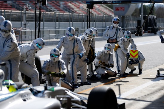
Both scenarios require a team of people to work seamlessly in a time critical and space-limited environment. In Formula One, a pit crew can change all four tyres on a car in around two seconds, with a team of nearly 20 people working in unison to successfully service a car. Williams has a dedicated human performance specialist who works with its pitcrew to fine tune the technique, processes, team work and health and fitness of team members.

Their experience previously treating new-borns in clinical practice has facilitated the transition of knowledge between the two industries and they have been the primary advisor to the hospital. Williams’s pitstops have been a real success story for the team in 2016, recording the fastest stops of any team at each of the first four races of the 2016 Formula One season.

Following these site visits, the neonatal team has identified and started implementing a number of changes to improve its resuscitation processes that are based on those used in Formula One racing. The resuscitation equipment trolley has now been audited and streamlined to ensure that equipment can be located as quickly as possible.

The neonatal team has mapped out a standardised floor space in delivery theatres to clearly show the area for the neonatal resuscitation team to work in; copying the customised floor map the Williams team takes to races to map out the specific pit box requirements at each track.

The pitstop resuscitation team at UHW are also in the early stages of implementing Formula One communications and analysis techniques, including the use of a “radio-check” prior to a resuscitation, greater use of hand signals rather than verbal communication, and video analysis to analyse performance following a resuscitation with debrief meetings as standard.

Speaking about the project Dr Rachel Hayward, specialist registrar in Neonates at the University Hospital of Wales said: “Resuscitation of a compromised neonate at delivery is time critical, requiring the provision of efficient and effective resuscitation to ensure an optimal outcome.”

Lovely the language medics use…

“Delays in providing effective resuscitative care can have marked consequences on survival or the development of long term complications. There is a growing amount of evidence to support a systematic approach to resuscitative care which is time-critical and dependent upon optimal team dynamics and clear communication.

“Analogous with the requirements of an effective pitstop we have worked with the Williams team to implement Formula One techniques and processes to augment neonatal resuscitative care”.

Claire Williams, Deputy Team Principal of Williams, added: “When we were approached by the Neonatal team at the University Hospital of Wales last year to offer some advice we were delighted to assist. Their work is vitally important and the pressure they work under is difficult to comprehend; it’s a matter of life and death every day of the week.

“If some of the advice we have passed on helps to save a young life then this would have been an extremely worthy endeavour. We are increasingly finding that Formula One know-how and technology can have benefit to other industries and this is a great example.”

I think this is great. We should have many more cross-industry knowledge transfers like this.

Gene breakthrough

Zoologists and bioengineers from Trinity College Dublin have identified over 1,000 genes whose responses change markedly when embryos are not able to move freely in the womb.

The discovery will help scientists better understand how important tissues are programmed to develop in our bodies which could, in turn, suggest how stem cells can be primed for use in tissue engineering and regenerative therapies.

The collaborative research conducted in the School of Natural Sciences and Trinity Centre for Bioengineering is addressing how embryonic movement influences bone and joint development. This research also furthers understanding of the consequences of reduced movement and shows how we might guide desired differentiation of bone and cartilage from stem cells.

Says Developmental Biologist and Associate Professor in Zoology, Paula Murphy who is the study’s senior author of the study:

“Why do babies move about so much while they are developing in the womb, particularly flexing their arms and legs? We know that if they don’t move enough, they are born with skeletal problems such as thin, fragile bones.

“Highly regulated signalling systems are needed for Mother Nature to follow the complex ‘recipes’ of genetic expression that enable the development of normal skeletons. What often surprises people is that mechanical signals also feed in to these signalling systems, and it is the movement of an embryo that sparks these.”

By studying how animals move and develop, the zoologists and bioengineers have pinpointed which steps during skeleton formation require stimulation from movement. Additionally, by examining the patterns of all the genes in the genome, they have shown which specific genes and molecules are stimulated by movement.

Contracting muscles and cartilage rudiments of the skeleton develop at the same time in the embryo, and the movement of the muscles influences normal development of bone and cartilage. Highlighted in this 3D image is the developing limb skeleton (left), muscle (right, in orange) and tendon (right, in green).

Contracting muscles and cartilage rudiments of the skeleton develop at the same time in the embryo, and the movement of the muscles influences normal development of bone and cartilage. Highlighted in this 3D image is the developing limb skeleton (left), muscle (right, in orange) and tendon (right, in green).

Very little is known about how the mechanical signals are integrated into the biochemical signalling pathways. This could soon change, however, as these researchers home in on the 1,000-plus genes whose responses changed in mouse embryos that lacked muscles and therefore did not kick during development.

The research, just published in the BMC Genomics journal, featured Research Fellow at Trinity, Rebecca Rolfe, as the first author. It highlighted a number of genes already known to encode regulatory molecules that guide developmental decisions in the embryo.

It also highlighted genes that are involved in controlling cell shape changes and in aiding cell-to-cell communication. In particular, the research highlighted the ‘Wnt’ pathway which passes signals from the exterior to the interior of specific cells, as a potential point of integration of mechanical and molecular signalling.

Adds Associate Professor Murphy:

“If we can better understand the signalling processes involved, we might guide development of stable bone and cartilage tissues for use in regenerative therapies. We are now working to fill in the gaps in our knowledge around the combinations of mechanical and molecular signals that are needed to guide differentiation of stem cells for this purpose.”

Spotting prostate cancer sooner

South west London’s St Anthony’s Hospital has introduced a new effective scan fusion procedure to help identify prostate cancer earlier than conventional methods.

A man’s risk of having prostate cancer is based on their PSA (protein prostate specific antigen) which is produced by the prostate gland.

This means that a scan and a biopsy need to be taken. The new technique called the BiopSee combines two diagnostic tools – an ultrasound scan with an MRI scan. This targets suspected cancerous areas of the prostate.

Stephen Gordon, St Anthony's Hospital

Stephen Gordon, St Anthony’s Hospital


In addition the samples can be taken from any part of the prostate and by avoiding the rectum, the risk of infection is reduced.

Stephen Gordon, Consultant Urological Surgeon, St Anthony’s Hospital, who introduced the purpose built fusion device to the hospital, says:

“The benefit of this approach is that not only is the prostate cancer detection rate likely to be better but that if cancer is found by targeting, then it is more likely to be of a significant nature which is best detected at an early stage. The procedure is performed as a day case under general anaesthetic.”

A standard biopsy is performed with an ultrasound probe in the back passage with the biopsy needle passing through the rectum into the prostate. On average 12 samples are taken randomly from different areas, which confirms prostate cancer about 35% of the time.

Adds Gordon:

“The problem with this is the random nature of the sampling and the fact that biopsies are taken via the back passage. This means the front or lower parts of the prostate are missed and despite being given antibiotics, men still suffer from infections.”

Prostate cancer is the most common cancer in men in the UK with 40,000+ men diagnosed with the disease and over a quarter of a million men living with the condition. Prostate cancer primarily affects older men (65 to 79) although 25% of cases occur in younger men.

Don’t forget the vitamin D

Our vitamin D levels will plummet this winter.

Vitamin D is a pro-hormone which our bodies naturally produce by synthesising ultraviolet light from the sun. It is needed for the body to make proper use of calcium for stronger bones and teeth and maintain a healthy immune system.

Ninety per cent of our essential vitamin D must come from our skin’s unprotected exposure to the sun, with only 10 per cent gained through diet. But the UK is currently in its vitamin D winter where winter sunshine is too weak to stimulate production of vitamin D.

When the sun is below 45 degrees in the sky, nearly all of the UVB it produces is absorbed by the atmosphere, rather than our bodies. So in the UK, the angle of our winter sun is simply not strong enough to ensure sufficient vitamin D production at the levels we need.

And even when the strength and angle of the sun is good enough for optimum vitamin D generation in our bodies, our nation’s infamous cloudy weather often blocks it out completely, hindering the absorption of those all important UVB rays. Therefore only a very few people are likely to generate enough vitamin D through natural sunlight exposure alone.

Diseases related to vitamin D deficiency, including multiple sclerosis, Type 2 diabetes, bone diseases such as osteoporosis and a growing number of internal cancers are believed to cost the UK taxpayer £29 billion annually – more than a quarter of the NHS budget.

More commonly, general symptoms associated with vitamin D deficiency include lack of energy and fatigue, headaches, poor immune system, muscle aches and depression.

So get out there and get yourself some supplementation advice. Vitamin D home test kits retail at £25.

Sniffing out stomach bugs could save NHS millions

Testing has begun on a device that can sniff out the presence of disease by smell.

Developed by Professors Chris Probert from the University of Bristol and Norman Ratcliffe from the University of the West of England, OdoReader uses pioneering technology to rapidly diagnose Clostridium difficile, by ‘reading’ the odour of stool samples. Clostridium difficile may cause severe diarrhoea, especially amongst hospital patients.

With the help of University Hospitals Bristol NHS Foundation Trust, the technology enables gasses emitted from faeces to be analysed in under an hour, leading to a rapid and inexpensive diagnosis. Such early detection could reap real health benefits for millions of people and help prevent the spread of infectious disease.

Gastro-intestinal diseases afflict over four billion adults and children each year. Delays in diagnosis can lead to patients being ill for longer, some may die, many will cost more to treat and infections may spread to other people. In England and Wales there are over 50,000 cases of Clostridium difficile reported each year: this infection prolongs a stay in hospital, is associated with a high number of deaths and costs the NHS £200 million annually.

The OdoReader device

A £1.3 million Wellcome Trust Translation Award will cover a three-year programme of work. It will support the development of OdoReader prototypes, which will then be tested against the industry gold standard method of making the diagnosis. The final produce will undergo a clinical trial before becoming available for sale around 2013. Says Chris Probert, Professor of Gastroenterology at the University of Bristol, and Consultant Gastroenterologist at University Hospitals Bristol:

“For a long time it has been known that stools have a distinctive and different odour if there is an infection. What OdoReader does is take this knowledge a step further by comparing the odour of faeces of patients with those from patients with specific gastro-intestinal disease to make a rapid diagnosis at the point of care.”

It’s also thought that OdoReader has potential far beyond that of being a portable device for diagnosing C difficile. It could be used for a range of other gastrointestinal disease as well as lung and urinary tract diseases.

%d bloggers like this: